

Cone Beam “CT Scan” with ULTRA LOW Dose radiation. Same-day CBCT scans on our Planmeca Viso G7, from full skull to C7 of the spine.

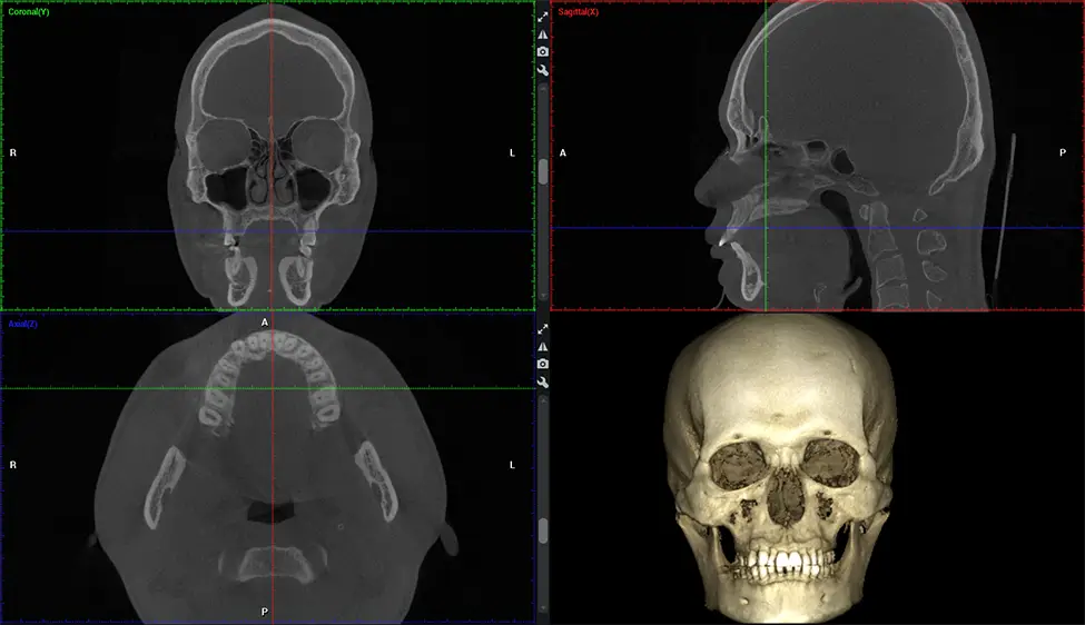
Cone Beam Computed Tomography (CBCT) is an advanced imaging technique used in dentistry and maxillofacial surgery to obtain detailed 3D images of the oral and maxillofacial structures.
Same Day CBCT Scan: $250.
*Interpretation – not included*
*Interpretation – not included*
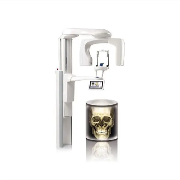
At Dr G Dental Studio, our CBCT scanners utilize a cone-shaped X-ray beam and a specialized detector to capture images from different angles. A computer then combines these images to create a 3D representation of the patient’s oral anatomy.
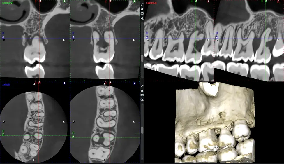
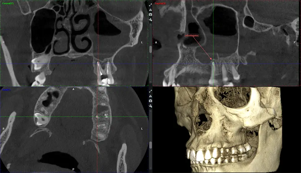
This 3D scan, called cone beam computed tomography, gives your dentist a more complete image of your oral anatomy and disease processes than a traditional X-ray. Unlike conventional X-rays, which capture a 2D image of your mouth from various angles, a 3D scan takes multiple digital X-rays for one image. It provides a complete view of your jaw, teeth, nerves, and soft tissues. This enhanced view allows dentists to detect minor issues not visible in traditional 2D scans, such as impacted wisdom teeth or bone fractures in the sinus cavity.
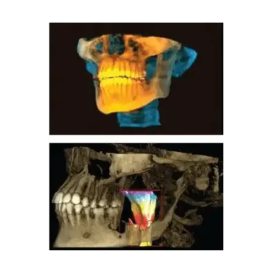
There are many benefits to using CBCT technology, especially compared to the traditional 2D X-ray format.
One of the most significant advantages of CBCT scans is that they provide much more information than traditional X-rays. A scan lets your dentist see images from all angles of your jaw and mouth, including your sinuses, nasal cavity, cheekbones, and other surrounding areas. This added information helps your dentist craft a comprehensive treatment plan that addresses all aspects of your oral health.
Another significant benefit is that 3D imaging provides more precise images of your bone structure. These images are more detailed, providing you with a more accurate diagnosis. An accurate diagnosis means better treatment for you.
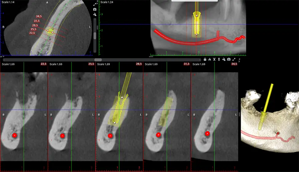
When paired with extractions, dental implant placement, or bone grafting, CBCT technology can provide you with a quicker and much more comfortable experience. This is because the 3D image provides a more detailed picture of your entire oral cavity, allowing your dentist to walk you through each step of the process before it occurs. This often helps patients feel more prepared and comfortable overall, especially if they have had a traumatic experience with past dental treatments.
The patient is first positioned in the CBCT scanner, which typically consists of a rotating arm that houses the X-ray source and a detector. The patient’s head is immobilized to ensure accurate image capture. The X-ray source and detector rotate around the patient’s head, capturing various X-ray images from multiple angles. As the X-ray source rotates, it emits the cone-shaped X-ray beam towards the detector. The detector captures the X-ray images, which are then processed by the CBCT software.
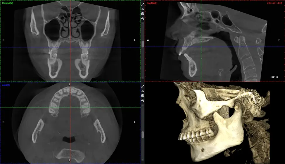
After the scanning process, the captured X-ray images are processed by the CBCT software, which applies algorithms to reconstruct a detailed 3D image of the scanned area. The software compiles these individual X-ray images and creates a digital 3D representation of the patient’s anatomy. The reconstructed 3D CBCT image can be viewed and analyzed by the dentist or radiologist. This image can be manipulated, rotated, and zoomed in or out to examine specific structures and evaluate the patient’s condition.
Complex oral surgeries, orthognathic (jaw) surgery, and removal of impacted teeth.
Benefits of CBCT (skull to C7)Offers precise, three-dimensional visualization of the skull, jaw, and craniocervical junction to enhance diagnostic accuracy and minimize surgical risks.
Assessing facial asymmetry, planning orthognathic surgery, and managing cleft lip and palate and sleep apnea.
Benefits of CBCT (skull to C7)Provides a comprehensive view of the craniofacial complex to better plan treatments and evaluate outcomes.
Diagnosing structural problems in the C1-C2 vertebrae, assessing misalignment and instability, and evaluating chronic pain.
Benefits of CBCT (skull to C7)Enables precise, weight-bearing views of the upper cervical spine, helping to identify abnormalities that inform targeted, non-invasive therapies.
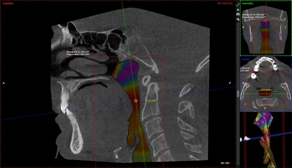
Diagnosing obstructive sleep apnea (OSA) and evaluating sinus and nasal cavity anatomy.
Benefits of CBCT (skull to C7)Visualizes the entire pharyngeal airway to measure volume, pinpoint constrictions, and plan corrective surgery if needed.
Screening and treating patients at risk for OSA.
Benefits of CBCT (skull to C7)Provides detailed 3D imaging for airway analysis, helping to create and adjust oral appliances and facilitate effective referrals to medical doctors.
Planmeca Viso G7 CBCT ( Cone Beam CT Scan ) is designed to surpass the demands of industry leaders, specialists, and large institutions. It has a large ø25×30 cm sensor with four built-in cameras. It can capture unlimited volume sizes from a ø3×3 cm to a ø30x30cm volume capturing the skullcap through C7 on the cervical spine. The Planmeca Viso G7 offers the industry’s largest single volume scan of ø30×19 cm. It’s poised to handle advanced imaging modalities such as Planmeca ProFace® and Planmeca 4D™ Jaw Motion technology. The occipital head support allows an unimpeded view of facial tissue.
Planmeca is the only manufacturer clinically proven to dramatically reduce patient radiation without sacrificing image quality. Planmeca Ultra Low Dose™ achieves an average reduction in dose of 77% without a statistical reduction in image quality allowing doctors to incorporate 3D imaging into routine protocols with effective patient radiation often lower than 2D intraoral imaging. This increases patient care, allows doctors to diagnose more dentistry, and often earlier in disease progression, than with 2D imaging alone.
Planmeca 3D units also include the industry’s only patient motion detection and correction technology, Planmeca CALM™. This technology corrects movement during CBCT acquisition reducing the risk of retakes.
Planmeca’s patented SCARA technology enables doctors to capture True Extraoral Bitewings that are as accurate at detecting caries as a 2D bitewing series, but with half the radiation dose. With these bitewings, doctors can view the apices of teeth on both jaws providing a more comprehensive view of the patients’ anatomy.
Planmeca 3D systems come with Autofocus, a technology that produces incredibly clear panoramic images, even on the interiors. This technology sets Planmeca’s 2D images apart from the competition.

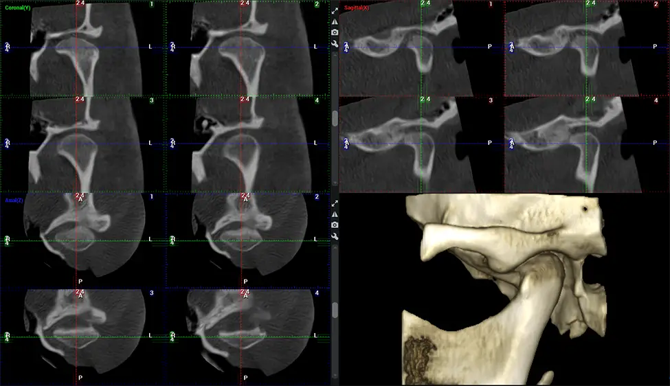

Same-day CBCT cone beam scans in Miami for $250 (interpretation not included). Call us to schedule.